

Client: Professor David Mazierski
Year: 2020
Media: Adobe Illustrator, Adobe Photoshop
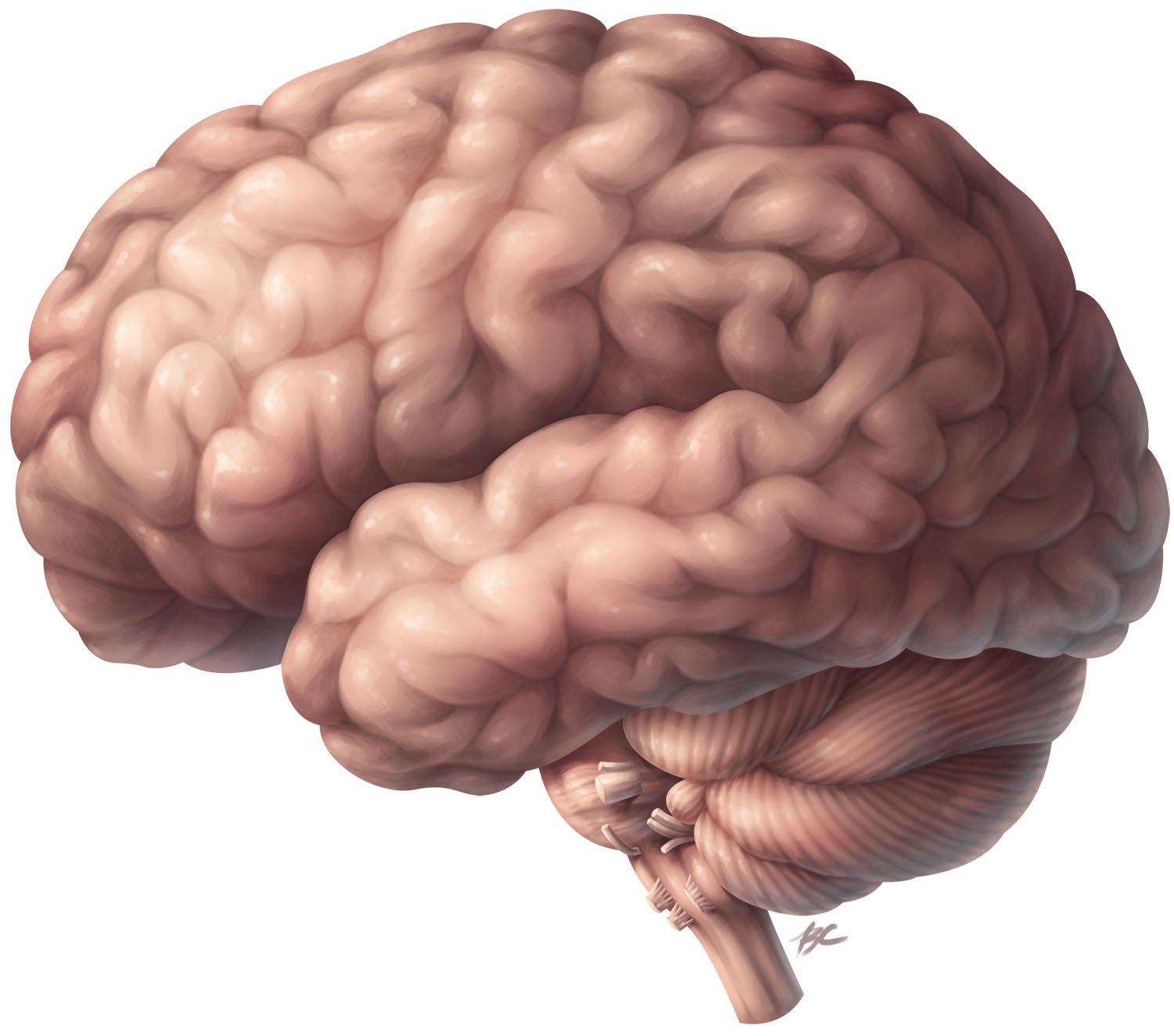
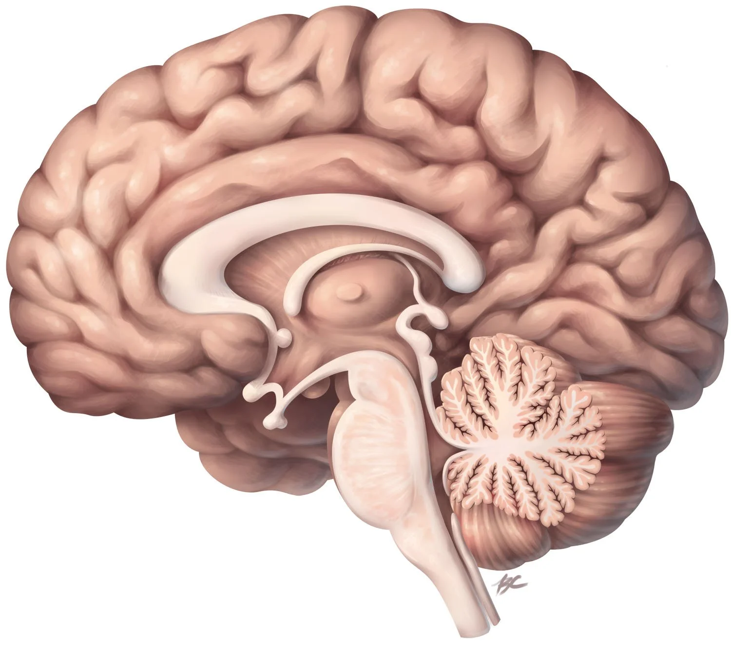
This piece was created to practice digital rendering and learn the detailed anatomy of the exterior and interior of the brain using a wide range of references to ensure accuracy.
References
Academic Sources
Agur, A. M. R., and Dalley, A. F. 2008. Grant’s Atlas of Anatomy, 12th ed. Philadelphia: Wolters Kluwer. Fig. 7.17: Middle meningeal artery and pterion, Fig. 7.20: Dura mater, and Fig. 7.21: Venous sinuses of the dura mater.
Gray, H. 2005. Gray’s Anatomy: The Anatomical Basis of Clinical Practice, 39th ed., ed. Standring, S. New York: Elsevier Churchill Livingstone. Fig 22.1: Lateral aspect of the left cerebral hemisphere indicating the major gyri and sulci, Fig. 22.2: Sagittal section of the brain, with the brain stem removed, showing the medial aspect of the left cerebral hemisphere, Fig. 22.3: Left lateral aspect of the brain, and Fig: 22.4: The medial surface of the left cerebral hemisphere after sagittal section of the brain, followed by removal of the brain stem and septum pellucidum.
Larsell, O. 1942. Anatomy of the Nervous System, 1st ed. New York: D. Appleton-Century Co. Fig. 269: Lateral view of the left cerebral hemisphere, and Fig. 271: Medial view of the brain.
Nieuwenhuys, R., Voogd, J., and van Huijzen, C. 2008. The Human Central Nervous System, 4th ed. Berlin: Springer. Fig. 3.2: Lateral view of the brain (1/1x), Fig. 3.7, Medial view of the right half of the bisected brain (1/1x), and Fig. 3.8: Medial view of the bisected brain stem and cerebellum (3/2x).
Schuenke, M., Schulte, E., and Schumacher, U. 2007. Thieme Atlas of Anatomy: Head and Neuroanatomy. Stuttgart, Germany: Thieme. Figre 5.3A: The four parts of the diencephalon, Figure 6.1Ec: Brainstem: Left lateral view, Figure 7.1B: Relationship of the cerebellum to the brainstem, and Figure 10.12B: Sagittal sections: VII and VIII (medial): Principal structures in the serial structures.